Experimental Laboratory Exercises
|
|
Experimental Laboratory Exercises | |
|
|
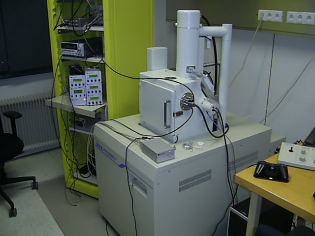
Scanning Electron MicroscopeA scanning electron microscope (SEM) uses a beam of high energy electrons to make images. The electrons are typically accelerated in the column to an energy of a few tens of keV and then focused by magnetic or electrostatic lenses into a narrow beam a few nanometers in diameter. Using a SEM it is possible to make images with higher resolution than with an optical microscope. Optical microscopes are typically limited by diffraction to a resolution about equal to a wavelength of light (about 1 micron). The resolution of a SEM is typically about one nanometer and not limited by diffraction since the wavelength of the electrons is typically much smaller than one nanometer.
Secondary electron imageThe most common to make an image in a SEM is to measure the secondary electrons that emitted by the specimen. The primary beam of high energy electrons enters the specimen and the electrons scatter inelastically with the other electrons. This transfers energy to the electrons in the specimen. Typically the electrons travel only a few nanometers before scattering. This results in a cloud of energetic electrons concentrated around the point where the beam enters the specimen. The figure below shows a simulation of the paths followed by the electrons.
Some of the energetic electrons within a scattering length of the surface of the specimen have enough energy to escape and are collected by the secondary electron detector. Vertical steps in the sample are clearly seen because when the electron beam is parallel to a surface, many energetic electrons are near the surface and the number of secondary electrons that escape the increases. The secondary electron current can be greater than the beam current in this case. If the secondary electrons are traveling in a direction that allows them to be captured by the secondary electron detector, the edge appear bright. If the secondary electrons are emitted in a direction away from the detector, the edge appears dark. This results in an image of the specimen that looks like it is illuminated by light from one side.
|